
ℹ️ What is Multiple Myeloma (MM)?
💊 Primary Treatment Approaches
Treatment for myeloma involves induction, possible stem cell transplant, and long-term maintenance — with novel immunotherapies transforming outcomes at relapse:
🔶 Plasma Cells & The Immune System — In Depth
To understand Multiple Myeloma, it helps to know where plasma cells come from and what they do. Plasma cells are fully differentiated B-lymphocytes — the final stage of B-cell maturation. Their sole purpose is to produce antibodies (immunoglobulins) that protect us from infections.
The Journey from B-Cell to Plasma Cell
When a B-cell encounters its target antigen, it undergoes activation and a remarkable transformation in the germinal center of lymph nodes:
Germinal Center Reactions
Critical processes that shape the antibody response:
- • Somatic Hypermutation (SHM) — random mutations in antibody genes to improve binding affinity
- • Class Switch Recombination (CSR) — switching from IgM to IgG, IgA, or IgE for specialized functions
- • Affinity Selection — only B-cells with the best-fitting antibodies survive
- • Clonal Expansion — selected B-cells multiply extensively
Terminal Differentiation
B-cells that exit the germinal center can become:
- • Short-lived Plasma Cells — produce antibodies for days to weeks in lymph nodes and spleen
- • Long-lived Plasma Cells — migrate to bone marrow; produce antibodies for years (even lifelong)
- • Memory B-cells — persist in lymphoid tissue for rapid response upon re-exposure
Myeloma arises from long-lived plasma cells in the bone marrow — explaining its marrow-centric biology.
Immunoglobulin Structure
Understanding antibody structure is essential because myeloma cells produce abnormal immunoglobulins — the monoclonal protein (M-protein) that is a hallmark of the disease:
| Ig Class | Heavy Chain | % of Myeloma | Key Features |
|---|---|---|---|
| IgG | γ (gamma) | ~50-55% | Most common myeloma subtype; most abundant Ig in blood |
| IgA | α (alpha) | ~20-25% | Second most common; dominant in mucosal immunity |
| Light Chain Only | κ or λ | ~15-20% | Only free light chains produced; higher risk of kidney damage |
| IgD, IgE, IgM | δ, ε, μ | <5% | Rare subtypes; IgM may overlap with Waldenström's macroglobulinemia |
💡 The M-protein (monoclonal protein or "M-spike" on electrophoresis) is produced by the myeloma clone. Tracking its level is a primary way to monitor disease burden and response to treatment.
📊 The Myeloma Spectrum: MGUS → SMM → Active Myeloma
Multiple Myeloma almost always evolves through a well-defined precursor pathway. Understanding this spectrum is critical because it creates opportunities for early detection and potentially interceptive therapy before full myeloma develops.
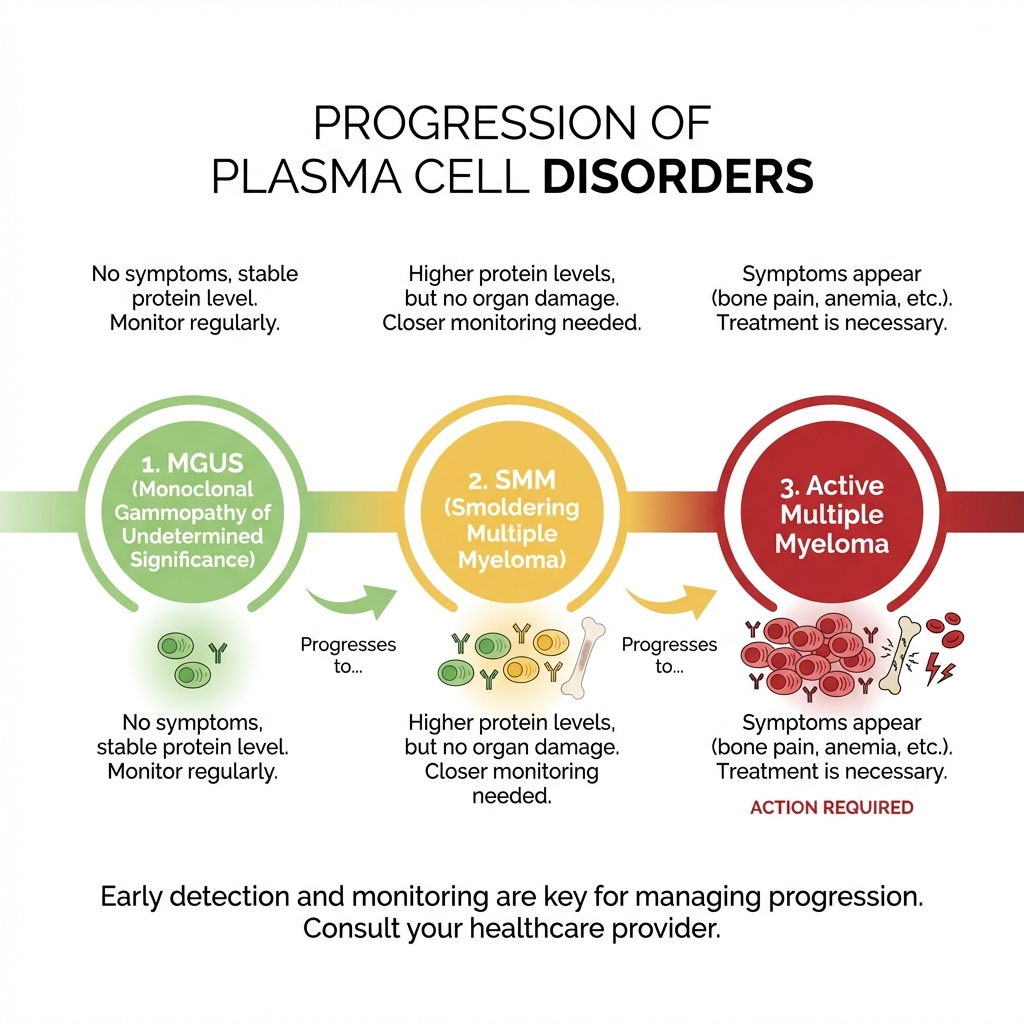
Figure 1: The spectrum of plasma cell disorders, from pre-malignant MGUS to active Multiple Myeloma.
MGUS — Monoclonal Gammopathy of Undetermined Significance
A pre-malignant condition present in ~3-4% of adults over age 50. The clonal plasma cells are present but not yet causing harm.
- • M-protein <3 g/dL
- • Bone marrow plasma cells <10%
- • No CRAB criteria or myeloma-defining events
Progression Risk:
~1% per year progress to myeloma or related malignancy. Lifelong monitoring is recommended — typically annual labs (SPEP, CBC, creatinine, calcium).
SMM — Smoldering Multiple Myeloma
An intermediate stage with higher tumor burden than MGUS but still no organ damage. Growing evidence supports early intervention for high-risk SMM.
- • M-protein ≥3 g/dL and/or
- • Bone marrow plasma cells 10-59%
- • No CRAB criteria or myeloma-defining events
Progression Risk:
~10% per year for the first 5 years, then ~3% per year. High-risk SMM (≥20% risk of progression within 2 years) may benefit from early treatment — active clinical trials are ongoing.
Active Multiple Myeloma — Requires Treatment
Treatment is initiated when myeloma causes organ damage (CRAB) or meets myeloma-defining events (MDEs).
CRAB Criteria:
- • Calcium elevation (>11 mg/dL)
- • Renal insufficiency (creatinine >2 mg/dL)
- • Anemia (hemoglobin <10 g/dL)
- • Bone disease (lytic lesions, osteoporosis, fractures)
Myeloma-Defining Events (since 2014):
- • Bone marrow plasma cells ≥60%
- • Serum free light chain ratio ≥100
- • >1 focal lesion on MRI (≥5mm each)
These MDEs indicate ultra-high-risk SMM that should be treated as active myeloma.
🦴 How Myeloma Damages the Body
Unlike leukemia, which primarily disrupts blood cell production, myeloma causes damage through multiple distinct mechanisms. The abnormal plasma cells and their products (M-protein and free light chains) cause harm to bones, kidneys, and the immune system simultaneously.
🦴 Bone Disease
The most characteristic feature of myeloma. Myeloma cells secrete factors (RANKL, DKK1, MIP-1α) that activate osteoclasts (bone-resorbing cells) while suppressing osteoblasts(bone-building cells). This creates lytic lesions — holes in the bone that cause pain, fractures, and spinal cord compression. Unlike other cancers with bone metastases, myeloma bone lesions do not heal even after successful treatment, because osteoblast suppression persists.
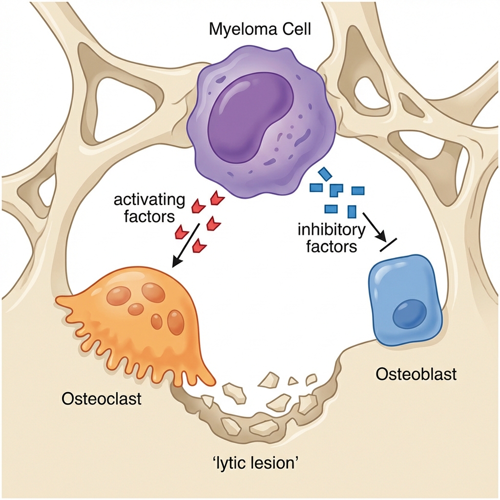
Figure 2: The imbalance between osteoclasts (bone-eating) and osteoblasts (bone-building) driven by myeloma cell signaling, leading to lytic lesions.
🫘 Kidney Damage
Affects ~20-50% of myeloma patients at diagnosis. The primary mechanism is cast nephropathy(\"myeloma kidney\"): excess free light chains form casts that obstruct kidney tubules. Other mechanisms include light chain deposition disease, amyloidosis (AL type), and hypercalcemia-induced kidney injury. Rapid reduction of light chains is critical to preserve kidney function.
🛡️ Immune Suppression
Myeloma causes profound immunoparesis — suppression of normal (polyclonal) immunoglobulin production. While the myeloma clone produces large amounts of a single, non-functional antibody, the levels of normal IgG, IgA, and IgM all decrease. This leaves patients highly susceptible to infections, particularly pneumonia (encapsulated bacteria like Streptococcus pneumoniae). Infection is the leading cause of early death in myeloma.
🩸 Hyperviscosity & Anemia
High M-protein levels can thicken the blood (hyperviscosity syndrome), causing headaches, blurred vision, and bleeding — most common in IgA and IgM myeloma. Myeloma also causes anemia through marrow infiltration, cytokine-mediated suppression of erythropoiesis, and kidney-related EPO deficiency. Fatigue from anemia is often the most impactful symptom on daily life.
⬆️ Hypercalcemia
As bones break down, large amounts of calcium are released into the bloodstream. High calcium levels cause confusion, excessive thirst, nausea, constipation, and kidney damage. Severe hypercalcemia is a medical emergency requiring immediate treatment with IV fluids, bisphosphonates, and calcitonin.
📋 Myeloma Genetics & Risk Stratification
Myeloma genetics are uniquely complex. Nearly all myeloma has chromosomal abnormalities, falling into two broad categories: hyperdiploid (extra chromosomes, generally better prognosis) and non-hyperdiploid (chromosomal translocations involving the immunoglobulin heavy chain locus on chromosome 14). Risk is stratified using FISH cytogenetics.
Primary Cytogenetic Abnormalities
These are the initiating events that drive myeloma development — present from the MGUS/SMM stage and retained throughout the disease course:
Hyperdiploidy (Extra Chromosomes)
Found in ~45-50% of myeloma. Cells have 48-75 chromosomes, typically with extra odd-numbered chromosomes (3, 5, 7, 9, 11, 15, 19, 21). Generally associated with standard-risk disease and better outcomes with modern therapies. However, some hyperdiploid subsets with concurrent high-risk features behave more aggressively.
t(11;14) — CCND1/IGH
Found in ~15-20% of myeloma. Upregulates cyclin D1, driving cell cycle progression. Generally standard risk. Notably, these myelomas are often BCL-2 dependent, making them uniquely sensitive to venetoclax — an oral BCL-2 inhibitor originally developed for CLL.
t(4;14) — MMSET/FGFR3/IGH
Found in ~12-15% of myeloma. Activates MMSET (an epigenetic modifier) and sometimes FGFR3. Historically associated with poor outcomes, but proteasome inhibitor-based regimens (bortezomib) have significantly improved prognosis. Now considered intermediate-to-high risk with modern treatment.
t(14;16) and t(14;20) — MAF Translocations
Each found in ~3-5% of myeloma. These translocations activate the MAF familyof transcription factors. Associated with adverse prognosis and aggressive disease biology.
Secondary/Progression Abnormalities
del(17p) — TP53 Deletion
Deletion of the short arm of chromosome 17, which contains TP53 — the \"guardian of the genome.\" Found in ~8-10% at diagnosis but increases at relapse. The single most adverse cytogenetic feature in myeloma, associated with chemoresistance, shorter remissions, and reduced overall survival. These patients typically require the most intensive approaches, often including transplant and novel combinations.
Gain(1q21) / del(1p)
Gain of 1q21 (amplification of chromosome 1q) is found in ~35-40% of myeloma and is increasingly recognized as an adverse risk factor, especially when >3 copies (amplification) are present. del(1p) is less common but also adverse. These chromosome 1 abnormalities are among the most common secondary events in myeloma progression.
📊 R-ISS Staging System
The Revised International Staging System (R-ISS) combines three factors to stratify myeloma prognosis: serum β2-microglobulin and albumin(reflecting tumor burden and patient fitness), LDH (reflecting tumor aggressiveness), and FISH cytogenetics (del(17p), t(4;14), t(14;16)). R-ISS Stage I has a 5-year overall survival of ~82%, Stage II ~62%, and Stage III ~40%.
🧬 The Bone Marrow Microenvironment
Myeloma is uniquely dependent on its bone marrow microenvironment. Unlike many cancers, myeloma cells cannot easily survive outside the marrow niche. Understanding this dependency has led to some of the most successful therapeutic strategies.
🏗️ Stromal Cell Support
Bone marrow stromal cells provide critical survival signals to myeloma cells through direct contact (adhesion molecules like VLA-4/VCAM-1) and secreted factors. This interaction activates NF-κB, JAK/STAT3, and PI3K/AKT pathways in myeloma cells, promoting survival and drug resistance. The stromal microenvironment also protects myeloma cells from chemotherapy-induced death — a phenomenon called cell adhesion-mediated drug resistance (CAM-DR).
🔄 The IL-6 Axis
Interleukin-6 (IL-6) is the dominant growth and survival factor for myeloma cells. Produced by stromal cells, osteoclasts, and myeloma cells themselves, IL-6 activates the JAK/STAT3 signaling cascade, driving proliferation, survival, and drug resistance. Early therapeutic attempts targeted IL-6 directly (siltuximab), but the most successful approach has been targeting downstream pathways through immunomodulatory drugs (IMiDs).
💊 Therapeutic Targeting of the Microenvironment
Many successful myeloma therapies work by disrupting the microenvironment interaction:
- • IMiDs (lenalidomide, pomalidomide) — degrade IKZF1/3 via cereblon, disrupting myeloma-stromal signaling
- • Proteasome inhibitors (bortezomib) — block NF-κB activation, a key survival pathway
- • Anti-CD38 (daratumumab) — directly targets myeloma cells via the ubiquitous CD38 surface marker
- • Bisphosphonates (zoledronic acid) — inhibit osteoclast-mediated bone destruction and may have anti-myeloma effects
💉 The Modern Treatment Landscape
Myeloma treatment has undergone a revolution over the past two decades. Median survival has improved from ~3 years in the 1990s to >8-10 years with modern therapy. While not yet routinely curable, deep and durable remissions are now achievable for many patients with combination regimens and novel immunotherapies.
📋 Treatment Phases
Induction
3-6 cycles of combination therapy to rapidly reduce disease burden. Standard regimens include VRd (bortezomib, lenalidomide, dexamethasone) ± daratumumab (Dara-VRd).
Consolidation
For eligible patients (typically age <70, good fitness): autologous stem cell transplant (ASCT) using high-dose melphalan. Deepens response achieved during induction.
Maintenance
Long-term therapy to maintain response. Lenalidomide maintenance (until progression) is standard of care, extending remission duration by years.
🎯 Breakthrough Immunotherapies (Relapsed/Refractory)
🧬 BCMA-Targeted CAR T-Cells
Idecabtagene vicleucel (Abecma®) and ciltacabtagene autoleucel (Carvykti®) target BCMA (B-cell maturation antigen) on myeloma cells. These have shown remarkable response rates (~70-98%) in heavily pre-treated patients. Carvykti® has moved to earlier lines of therapy based on extraordinary trial results.
💊 Bispecific Antibodies
Teclistamab (Tecvayli®) (BCMA×CD3), talquetamab (Talvey®) (GPRC5D×CD3), and elranatamab (Elrexfio®)(BCMA×CD3) redirect T-cells to kill myeloma cells. Available \"off the shelf\" unlike CAR T — no manufacturing wait time. Given as subcutaneous injections.
⚡ CELMoDs (Next-Gen IMiDs)
Mezigdomide and iberdomide are next-generation cereblon modulators designed to overcome resistance to lenalidomide and pomalidomide. They achieve deeper degradation of IKZF1/3, showing activity in IMiD-refractory patients.
📊 MRD in Myeloma
Minimal Residual Disease (MRD) negativity is emerging as the gold standard for treatment response in myeloma. Achieved when fewer than 1 myeloma cell per million bone marrow cells is detected (10⁻⁶ sensitivity via next-generation flow or sequencing). Patients who achieve sustained MRD negativity have significantly longer progression-free and overall survival, regardless of cytogenetic risk. MRD testing is increasingly used to guide treatment duration decisions, potentially allowing MRD-negative patients to stop therapy.
📚 Authoritative Myeloma Resources
These trusted organizations provide comprehensive, up-to-date information about Multiple Myeloma:
Blood Cancer United — Myeloma
Comprehensive myeloma overview, diagnosis, and treatment information.
Visit →IMF — International Myeloma Foundation
The leading myeloma-specific foundation: patient handbooks, InfoLine, and research updates.
Visit →NCI — Myeloma Treatment (PDQ®)
Detailed treatment information reviewed by oncology experts.
Visit →LLS — Multiple Myeloma
Patient guides, caregiver support, and financial assistance programs.
Visit →🛠️ Tools & Focused Research
Scientific Literature
Search thousands of PubMed articles curated for Multiple Myeloma.
Clinical Trials
Explore actively recruiting clinical trials for this disease group.
View Clinical Trials