
ℹ️ What is Acute Myeloid Leukemia (AML)?
💊 Primary Treatment Approaches
Treatment for AML depends on subtype, genetic mutations, patient age, and fitness. The main categories include:
🔶 The Myeloid Lineage — In Depth
To understand AML, it helps to know how normal myeloid blood cells develop. All blood cells originate from hematopoietic stem cells (HSCs) in the bone marrow. These stem cells first commit to one of two main lineages: myeloid or lymphoid. AML specifically affects the myeloid lineage.
The Differentiation Cascade
Myeloid differentiation follows a carefully regulated cascade. The HSC first becomes a Common Myeloid Progenitor (CMP), which then branches into two key pathways:
Granulocyte-Monocyte Progenitor (GMP)
Gives rise to the granulocytes and monocytes — key players in innate immunity:
- • Neutrophils — the most abundant white blood cells; first responders to bacterial infection
- • Monocytes / Macrophages — engulf pathogens and cellular debris; coordinate immune responses
- • Eosinophils — combat parasites and modulate allergic responses
- • Basophils / Mast cells — mediate inflammatory and allergic reactions
Megakaryocyte-Erythroid Progenitor (MEP)
Gives rise to the oxygen-carrying and clotting components of blood:
- • Erythrocytes (Red Blood Cells) — carry oxygen via hemoglobin; ~70% of all marrow output
- • Megakaryocytes → Platelets — giant cells that fragment into thousands of platelets for blood clotting
Growth Factors That Drive Differentiation
Each step of differentiation is guided by specific growth factors (also called cytokines). These chemical signals tell progenitor cells which type of blood cell to become:
| Growth Factor | Full Name | Drives Production Of |
|---|---|---|
| G-CSF | Granulocyte Colony-Stimulating Factor | Neutrophils |
| M-CSF | Macrophage Colony-Stimulating Factor | Monocytes / Macrophages |
| GM-CSF | Granulocyte-Macrophage CSF | Both granulocytes and monocytes |
| EPO | Erythropoietin | Red blood cells (erythrocytes) |
| TPO | Thrombopoietin | Platelets (via megakaryocytes) |
💡 G-CSF is commonly given to patients after chemotherapy (as Neupogen® or Neulasta®) to help neutrophil counts recover faster.
🔬 What Are Blast Cells?
The word "blast" comes from the Greek blastos meaning "germ" or "bud." In hematology, blasts are immature precursor cells — the earliest identifiable cells in the differentiation cascade that will eventually mature into functional blood cells.
Normal vs. Leukemic Blasts
✅ Normal Blasts
- • Comprise <5% of bone marrow cells
- • Rapidly differentiate into mature, functional cells
- • Respond normally to growth factor signals
- • Undergo programmed cell death (apoptosis) when not needed
⚠️ Leukemic Blasts (Myeloblasts)
- • Accumulate to ≥20% of marrow (the diagnostic threshold for AML)
- • Maturation arrest: they proliferate but fail to differentiate
- • Ignore normal growth signals; proliferate unchecked
- • Resist apoptosis, allowing them to accumulate
How Blasts Are Identified
Pathologists identify blasts using multiple complementary techniques:
🔎 Morphology (Under the Microscope)
Blasts have a characteristic appearance: large nucleus relative to cell size, fine chromatin (open, less condensed DNA), prominent nucleoli (1-4 visible), and scant basophilic cytoplasm (thin rim of blue-staining cell body).
💎 Auer Rods
A hallmark finding in AML: Auer rods are needle-like crystalline structures within the cytoplasm of leukemic blasts. They are aggregates of azurophilic granules and are pathognomonic for myeloid lineage — meaning if Auer rods are present, the leukemia is definitively myeloid, not lymphoid.
🧪 Flow Cytometry (Immunophenotyping)
Flow cytometry identifies cell surface markers to confirm blast identity. Key myeloid blast markers include: CD34 (stem/progenitor marker), CD117 (c-KIT), HLA-DR, CD13, CD33, and MPO (myeloperoxidase). The specific pattern helps distinguish AML from ALL and identify AML subtypes.
📊 The 20% Threshold
The WHO and ICC classifications define AML when blast percentage reaches ≥20% in the bone marrow or peripheral blood. However, certain genetic abnormalities (like PML::RARA in APL, or RUNX1::RUNX1T1) qualify as AML regardless of blast count — these are called "AML-defining genetic abnormalities."
🦴 Normal Bone Marrow vs. AML Bone Marrow
The bone marrow is a finely balanced ecosystem. In AML, this balance is catastrophically disrupted. Understanding the difference between healthy and leukemic marrow illustrates why AML causes the symptoms it does.
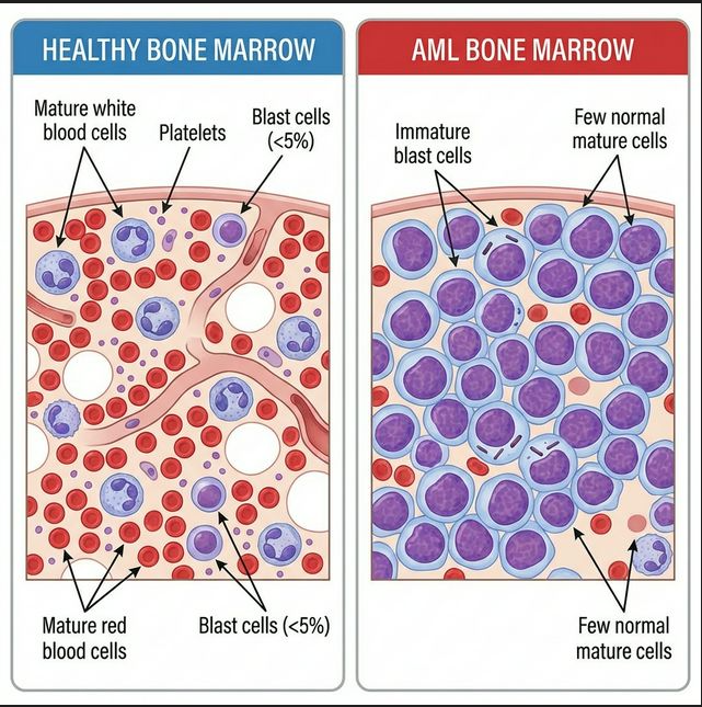
In AML, leukemic blasts overwhelm the marrow, crowding out normal cells and causing the hallmark complications: anemia, infections, and bleeding.
How AML Disrupts Normal Blood Production
🛑 Maturation Arrest
The defining feature of AML: leukemic cells are frozen at an immature stage. They continue to proliferate but cannot mature into functional blood cells. The specific stage of arrest determines the AML subtype (e.g., promyelocytic in APL, monoblastic in AML-M5).
📦 The Crowding Effect
Leukemic blasts don't just fail to work — they physically displace normal hematopoietic cells. The marrow becomes "packed" with blasts (often >80-90% cellularity), leaving little room for normal red cell, white cell, and platelet production.
🩸 Resulting Cytopenias
The crowding effect produces the classic triad of AML symptoms: anemia (fatigue, pallor, shortness of breath from low red cells), neutropenia (infections from low functional white cells), and thrombocytopenia (bleeding, bruising from low platelets).
🌊 Extramedullary Disease
In some cases, blasts spill out of the marrow into the peripheral blood (leading to very high WBC counts called hyperleukocytosis). Blasts can also infiltrate other organs — the spleen, liver, gums, skin, or central nervous system — causing organ-specific symptoms. Collections of blasts outside the marrow are called myeloid sarcomas (or chloromas).
📋 AML Classification
AML is not a single disease — it encompasses many subtypes with distinct biology, behavior, and treatment responses. Modern classification systems group AML by its genetic driversrather than just how cells look under the microscope.
WHO 2022 / ICC Classification
The current standard classifiers (WHO 5th edition and ICC 2022) prioritize genetic abnormalities as the primary basis for classification:
AML with Recurrent Genetic Abnormalities
Defined by specific chromosomal translocations or gene mutations. Examples: PML::RARA (APL), RUNX1::RUNX1T1, CBFB::MYH11 (core-binding factor AML), KMT2A rearrangements, NPM1-mutated AML, CEBPA-mutated AML. These often have specific treatment implications and prognosis.
AML with Myelodysplasia-Related Changes
AML that evolved from MDS or has MDS-like features (multi-lineage dysplasia, MDS-related cytogenetic changes). Often presents in older patients and may be more resistant to standard chemotherapy. CPX-351 (Vyxeos®) was specifically approved for this subtype.
Therapy-Related AML (t-AML)
AML that develops after prior chemotherapy or radiation for another cancer. Accounts for ~10% of AML cases. Often carries adverse-risk genetics (TP53 mutations, complex karyotype).
AML, Not Otherwise Specified (AML-NOS)
AML that doesn't fit into the above genetic categories. This is where the legacy FAB classification (M0 through M7) is still sometimes referenced to describe the morphological appearance and lineage of the blasts.
📜 FAB Classification (Historical Reference)
The French-American-British (FAB) system, developed in the 1970s, classified AML based on cell morphology and differentiation. While largely replaced by genetic classification, these designations are still commonly used:
Minimally differentiated
Without maturation
With maturation
Promyelocytic (APL)
Myelomonocytic
Monocytic
Erythroid
Megakaryoblastic
🧬 Key AML Mutations — In Context
AML is driven by combinations of mutations, not single genes acting alone. Understanding how mutations interact and co-occur is essential for interpreting your molecular report and understanding your treatment plan.
Founding vs. Cooperating Mutations
AML typically develops through a multi-hit model: an initial "founding" mutation creates a pre-leukemic state, and additional "cooperating" mutations drive transformation to full AML:
🏗️ Founding Mutations
Early events that create a clonal expansion. Examples: DNMT3A, TET2, ASXL1, IDH1/2. These are often found in clonal hematopoiesis of indeterminate potential (CHIP) — a pre-malignant state that can exist years before AML develops.
⚡ Cooperating Mutations
Later events that push the clone toward AML. Examples: FLT3-ITD, NPM1, RAS pathway mutations. These typically affect signaling pathways that drive proliferation or block differentiation.
Important Mutation Profiles
NPM1-Mutated AML
Present in ~30% of AML cases. NPM1 mutations cause the nucleophosmin protein to mislocalize to the cytoplasm. Associated with favorable prognosis when present without FLT3-ITD. Responds well to standard chemotherapy; transplant in first remission may not be needed.
Common co-occurrence: DNMT3A + NPM1 + FLT3 ("triple mutant") — prognosis depends on FLT3-ITD allelic ratio.
FLT3 Mutations (ITD and TKD)
FLT3-ITD (internal tandem duplication) is found in ~25% of AML and drives aggressive proliferation via constitutive activation of the FLT3 receptor tyrosine kinase. FLT3-TKD (tyrosine kinase domain point mutations) confers less adverse risk.
Targeted therapies: midostaurin (added to induction), gilteritinib (relapsed/refractory). ELN 2022 no longer uses the FLT3-ITD allelic ratio for risk stratification.
IDH1 and IDH2 Mutations
Isocitrate dehydrogenase mutations (IDH1 ~8%, IDH2 ~12% of AML) produce an abnormal metabolite called 2-hydroxyglutarate (2-HG) that blocks cell differentiation. This gives them a unique therapeutic vulnerability.
Targeted therapies: ivosidenib (IDH1), enasidenib (IDH2). These drugs can induce differentiation of leukemic blasts, sometimes achieving remission without intensive chemotherapy.
TP53 Mutations
The "guardian of the genome" — TP53 normally prevents damaged cells from surviving. Mutations (~5-10% of de novo AML, higher in therapy-related AML) are associated with complex karyotype, chemotherapy resistance, and the worst prognosis of any AML subgroup.
Active research areas: magrolimab (CD47 antibody), eprenetapopt (APR-246), and other novel agents specifically targeting TP53-mutated AML.
CEBPA Mutations
CEBPA encodes a transcription factor essential for granulocyte differentiation. Biallelic CEBPA mutations (or bZIP in-frame mutations per ELN 2022) are associated with favorable prognosis. These patients often respond well to chemotherapy alone.
DNMT3A, TET2, and ASXL1
These epigenetic regulators are among the most commonly mutated genes in AML. They affect how DNA is read (methylation and chromatin modification) without changing the DNA sequence itself.
- • DNMT3A R882 — most common AML mutation overall; often one of the earliest events
- • TET2 — involved in DNA demethylation; loss impairs differentiation
- • ASXL1 — chromatin modifier; associated with adverse prognosis (ELN 2022 adverse when mutated)
🔗 Connection to ELN 2022 Risk
The European LeukemiaNet (ELN) 2022 guidelines use mutation status as the primary basis for risk classification: Favorable (NPM1 without adverse markers, biallelic CEBPA, core-binding factor), Intermediate (NPM1 with certain co-mutations, wild-type NPM1 without adverse markers), and Adverse (TP53, ASXL1, RUNX1, MECOM rearrangements, complex/monosomal karyotype). This classification directly guides transplant decisions.
📚 Authoritative AML Resources
These trusted organizations provide comprehensive, up-to-date information about AML:
Blood Cancer United — AML
Comprehensive AML overview, diagnosis, and treatment information.
Visit →LLS — Acute Myeloid Leukemia
Patient guides, support programs, and clinical trial finder.
Visit →NCI — AML Treatment (PDQ®)
Detailed treatment information reviewed by oncology experts.
Visit →NCCN — AML Patient Guidelines
Patient-friendly version of the clinical practice guidelines.
Visit →🛠️ Tools & Focused Research
Scientific Literature
Search thousands of PubMed articles curated for Acute Myeloid Leukemia.
Clinical Trials
Explore actively recruiting clinical trials for this disease group.
View Clinical Trials