
ℹ️ What is Acute Lymphoblastic Leukemia (ALL)?
💊 Primary Treatment Approaches
Treatment for ALL spans 2-3 years and varies by age group, genetic subtype, and response to initial therapy (measured by MRD). Key approaches include:
🔷 The Lymphoid Lineage — In Depth
To understand ALL, it helps to know how normal lymphoid blood cells develop. All blood cells originate from hematopoietic stem cells (HSCs) in the bone marrow. These stem cells first commit to one of two main lineages: myeloid or lymphoid. ALL specifically affects the lymphoid lineage.
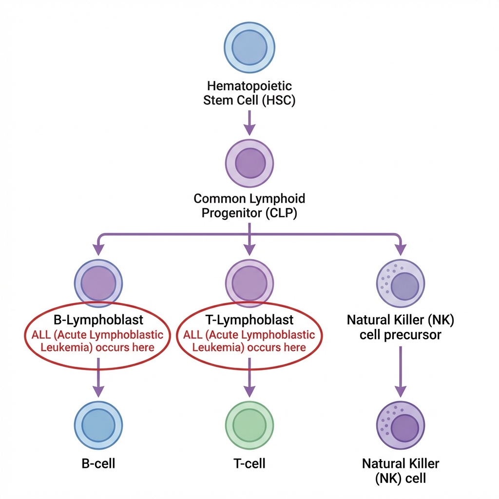
Figure 1: The lymphoid differentiation cascade. ALL occurs when lymphoblasts (precursors) fail to mature and begin multiplying uncontrollably.
The Lymphoid Differentiation Cascade
Lymphoid differentiation begins when the HSC becomes a Common Lymphoid Progenitor (CLP), which then branches into three key cell types essential for adaptive and innate immunity:
B-Cell Lineage
The most commonly affected lineage in ALL (~85% of cases):
- • Pro-B cells — earliest committed B-cell precursors; begin immunoglobulin gene rearrangement
- • Pre-B cells — express cytoplasmic μ heavy chain; undergoing light chain rearrangement
- • Immature B cells — express surface IgM; undergo self-tolerance testing
- • Mature B cells → Plasma cells — produce antibodies; key to humoral immunity
T-Cell Lineage
Accounts for ~15% of ALL cases; precursors migrate to the thymus for maturation:
- • Thymocytes — progenitors that undergo positive and negative selection in the thymus
- • CD4+ Helper T cells — coordinate immune responses by signaling other immune cells
- • CD8+ Cytotoxic T cells — directly kill virus-infected or cancerous cells
- • Regulatory T cells — prevent autoimmunity by suppressing excess immune activity
NK Cell Lineage
Natural Killer cells bridge innate and adaptive immunity:
- • NK cells — rapidly kill abnormal cells without prior sensitization
- • Tumor surveillance — recognize and destroy cells lacking MHC-I molecules
- • Viral defense — provide early defense before adaptive immunity activates
- • NK/T-cell ALL — rare but recognized subtype
Growth Factors That Drive Lymphoid Differentiation
Each step of lymphoid differentiation is guided by specific cytokines and growth factors. These signals tell progenitor cells which type of lymphocyte to become:
| Growth Factor | Full Name | Primary Role |
|---|---|---|
| IL-7 | Interleukin-7 | Essential for B-cell and T-cell development; survival signal for lymphoid progenitors |
| IL-2 | Interleukin-2 | T-cell growth and activation; promotes regulatory T-cell maintenance |
| IL-15 | Interleukin-15 | NK cell development and survival; memory T-cell maintenance |
| IL-3 | Interleukin-3 | Multi-lineage growth factor; supports early progenitor survival |
| SCF | Stem Cell Factor (c-KIT ligand) | Early lymphoid progenitor survival and proliferation |
💡 IL-7 signaling is particularly important in ALL biology — aberrant IL-7 receptor signaling is found in some T-ALL cases and is being explored as a therapeutic target.
🔬 What Are Lymphoblasts?
Lymphoblasts are immature lymphoid precursor cells — the earliest identifiable cells in the lymphoid differentiation cascade. In ALL, these cells are frozen at an immature stage and accumulate uncontrollably, unable to mature into functional lymphocytes.
Normal vs. Leukemic Lymphoblasts
✅ Normal Lymphoblasts
- • Comprise <5% of bone marrow cells
- • Rapidly differentiate into mature B-cells, T-cells, or NK cells
- • Respond to normal cytokine signals (IL-7, IL-2)
- • Undergo apoptosis when not needed or self-reactive
⚠️ Leukemic Lymphoblasts
- • Accumulate to ≥20-25% of marrow (the diagnostic threshold for ALL)
- • Maturation arrest: proliferate but fail to become functional lymphocytes
- • Acquire proliferative and survival advantages through genetic mutations
- • Resist apoptosis and evade normal immune surveillance
How Lymphoblasts Are Identified
Pathologists use multiple complementary techniques to identify and classify lymphoblasts:
🔎 Morphology (Under the Microscope)
Lymphoblasts are typically small to medium-sized cells with a high nuclear-to-cytoplasmic ratio, condensed chromatin, inconspicuous nucleoli, and scant agranular cytoplasm. Unlike myeloblasts, lymphoblasts lack Auer rods and are MPO-negative (myeloperoxidase negative) — a key distinguishing feature from AML.
🧪 Flow Cytometry (Immunophenotyping)
Flow cytometry is critical for classifying ALL as B-cell or T-cell lineage. Key markers include:
B-ALL Markers
CD19, CD22, CD79a, CD10 (CALLA), TdT, PAX5
T-ALL Markers
CD3 (cytoplasmic), CD7, CD5, CD2, CD1a, TdT
🧬 Cytogenetics & Molecular Testing
Chromosome analysis and molecular testing identify genetic abnormalities that determine subtype, prognosis, and treatment. FISH, karyotyping, and next-generation sequencing panels are standard at diagnosis to detect translocations like BCR-ABL1 (Philadelphia chromosome), ETV6-RUNX1, and KMT2A rearrangements.
📊 The 20-25% Blast Threshold
The WHO classification defines ALL when lymphoblast percentage reaches ≥20% in the bone marrow. Some protocols use a ≥25% threshold (particularly in pediatric oncology). However, certain genetic abnormalities — such as BCR-ABL1 with lymphoblast morphology — can support an ALL diagnosis even with lower blast counts when combined with immunophenotype and clinical context.
🦴 Normal Bone Marrow vs. ALL Bone Marrow
The bone marrow is a finely balanced ecosystem. In ALL, this balance is catastrophically disrupted as immature lymphoblasts overwhelm the marrow space. Understanding this disruption explains why ALL causes the symptoms it does — and why it can spread beyond the marrow.
How ALL Disrupts Normal Blood Production
🛑 Maturation Arrest
The defining feature of ALL: leukemic lymphoid cells are frozen at an immature stage. They continue to divide but cannot mature into functional B-cells or T-cells. The specific stage of arrest — pro-B, pre-B, or thymic — helps classify the ALL subtype and guide treatment.
📦 The Crowding Effect
Leukemic lymphoblasts physically displace normal hematopoietic cells. The marrow becomes packed with blasts (often >90% cellularity), leaving little room for normal red blood cell, white blood cell, and platelet production — leading to the classic symptoms of ALL.
🩸 Resulting Cytopenias
The crowding effect produces the classic triad of symptoms: anemia (fatigue, pallor, shortness of breath), neutropenia (recurrent infections from low functional white cells), and thrombocytopenia (bleeding, easy bruising from low platelets).
🧠 CNS Involvement
A critical difference from AML: ALL has a strong tendency to infiltrate the central nervous system (CNS). Lymphoblasts can cross the blood-brain barrier and accumulate in the cerebrospinal fluid, causing headaches, vision changes, and cranial nerve palsies. This is why intrathecal chemotherapy (injected directly into the spinal fluid) and sometimes cranial radiation are standard parts of ALL treatment protocols.
🫁 Extramedullary Disease
Beyond the CNS, lymphoblasts can infiltrate other organs. T-ALL frequently presents with a mediastinal (thymic) mass — an enlargement in the chest that can cause breathing difficulty. Both B-ALL and T-ALL can cause lymphadenopathy (swollen lymph nodes), hepatosplenomegaly (enlarged liver and spleen), and less commonly testicular involvement (a sanctuary site in males).
📋 ALL Classification
ALL is not a single disease — it encompasses many subtypes defined by the cell lineage (B or T), the stage of maturation arrest, and critically, the underlying genetic abnormalities. Modern classification integrates immunophenotype with genetics to guide treatment decisions.
WHO 2022 Classification: Major Categories
B-Lymphoblastic Leukemia/Lymphoma (~85% of ALL)
The most common form. Further classified by recurrent genetic abnormalities: BCR-ABL1 (Philadelphia+), ETV6-RUNX1 (most common in children), hyperdiploidy (>50 chromosomes, favorable in children), KMT2A-rearranged (common in infant ALL), TCF3-PBX1, BCR-ABL1-like (Ph-like), and others. The genetic subtype strongly influences prognosis and treatment intensity.
T-Lymphoblastic Leukemia/Lymphoma (~15% of ALL)
Arises from T-cell precursors, often in the thymus. More common in adolescents and young adult males. Frequently presents with a mediastinal mass and high white blood cell counts. Subtypes include Early T-precursor (ETP) ALL — which has a gene expression profile resembling myeloid cells and may benefit from different therapeutic approaches.
Childhood vs. Adult ALL
A critical distinction in ALL biology: the disease behaves very differently across age groups.
👶 Childhood ALL (Ages 1-9)
- • ~90% cure rate with modern protocols
- • More likely to have favorable genetics (hyperdiploidy, ETV6-RUNX1)
- • Most common cancer in children
- • Treatment duration: 2-3 years (includes maintenance phase)
- • Long-term survivorship concerns: growth, fertility, secondary cancers
🧑 Adult ALL (Ages 15+)
- • ~40-50% cure rate (improving with pediatric-inspired regimens)
- • More likely to have adverse genetics (BCR-ABL1, KMT2A, Ph-like)
- • Higher prevalence of T-ALL and Ph+ ALL
- • Adolescents/young adults (AYA, 15-39) benefit from pediatric protocols
- • Older adults often cannot tolerate intensive chemotherapy
📊 Maturation Stage Classification
B-ALL is further subclassified by the stage of B-cell development at which maturation arrest occurs. This is determined by immunophenotyping (flow cytometry):
CD19+, CD10−
Earliest B-cell stage
CD10+ (CALLA)
Most common subtype
Cytoplasmic μ+
Heavy chain expression
Surface Ig+
Burkitt-type / rare
🧬 Key ALL Genetic Abnormalities — In Context
Genetic abnormalities are the most important prognostic factors in ALL and increasingly guide treatment choices. Unlike AML, where mutations accumulate over time, ALL is more often driven by chromosomal translocations and copy number alterations that disrupt lymphoid development.
Prognostically Important Genetics
ETV6-RUNX1 (TEL-AML1) Translocation
The most common genetic abnormality in childhood B-ALL (~25% of pediatric cases). This translocation t(12;21) creates an abnormal fusion transcription factor. Associated with excellent prognosis in children with >95% cure rates. Rare in adult ALL.
Often associated with hyperdiploidy and age 2-9 years — the "good prognosis" triad of childhood ALL.
High Hyperdiploidy (>50 Chromosomes)
Found in ~25-30% of childhood B-ALL. Cells have 51-67 chromosomes (normal is 46), typically with extra copies of chromosomes 4, 10, 17, and 21. Associated with favorable prognosis, particularly when combined with age 1-9 years. Hyperdiploid cells are more susceptible to methotrexate and mercaptopurine therapy.
BCR-ABL1 (Philadelphia Chromosome)
The t(9;22) translocation creates the Philadelphia chromosome, producing a constitutively active tyrosine kinase. Found in ~3-5% of childhood ALL but ~25% of adult ALL — making it the most common genetic abnormality in adult ALL. Historically associated with very poor prognosis, but outcomes have dramatically improved with the addition of tyrosine kinase inhibitors.
Targeted therapies: imatinib, dasatinib, ponatinib. Adding TKIs to chemotherapy has transformed Ph+ ALL from one of the worst prognoses to near-comparable outcomes in many patients.
Ph-like (BCR-ABL1-like) ALL
Has a gene expression profile similar to Ph+ ALL but lacks the BCR-ABL1 fusion. Instead, these cases harbor diverse kinase-activating alterations (CRLF2, JAK2, ABL-class fusions, EPOR, etc.). Found in ~15-20% of B-ALL cases. Associated with poor prognosis with standard therapy, but many harbored fusions may respond to specific TKIs or JAK inhibitors.
Emerging targeted approaches: dasatinib (for ABL-class fusions), ruxolitinib (for JAK-STAT pathway alterations).
KMT2A (MLL) Rearrangements
Found in ~80% of infant ALL (under 1 year) and ~5-10% of adult ALL. The KMT2A gene (chromosome 11q23) is a histone methyltransferase involved in gene regulation. KMT2A-rearranged ALL has a distinct biology — often with a pro-B immunophenotype, high WBC count, and involvement of the CNS. Associated with poor outcomes, particularly in infants.
Active research: menin inhibitors (revumenib, ziftomenib) target the KMT2A-menin interaction and show promising results in clinical trials.
Hypodiploidy (<44 Chromosomes)
Near-haploid (24-31 chromosomes) and low-hypodiploid (32-39 chromosomes) ALL carry the worst prognosis of any ALL subtype. Often associated with TP53 mutations(especially in low-hypodiploid cases). These cases frequently require transplant in first remission.
IKZF1 Deletions (IKAROS)
IKZF1 encodes the IKAROS transcription factor, essential for normal lymphoid development. Deletions or mutations of IKZF1 are found in ~15% of pediatric and ~40% of adult B-ALL. They confer adverse prognosis independent of other risk factors and are a hallmark of Ph-like ALL. IKZF1 status is now used in risk stratification in multiple treatment protocols.
🎯 MRD: The Modern Risk Measure
Minimal Residual Disease (MRD) measurement has become the single most important prognostic factor in ALL, often superseding genetics. MRD quantifies the remaining leukemia cells after treatment (by flow cytometry or PCR), detecting as few as 1 leukemia cell among 10,000-1,000,000 normal cells. Patients who achieve MRD-negative status (<0.01%) after induction have significantly better outcomes. MRD status guides decisions about treatment intensification, transplant, and immunotherapy (e.g., blinatumomab for MRD+ patients).
💉 Immunotherapy Revolution in ALL
ALL has been at the forefront of the immunotherapy revolution. These breakthrough therapies have transformed outcomes for patients with relapsed or refractory disease and are increasingly being incorporated into frontline treatment.
🎯 Blinatumomab (Blincyto®)
A bispecific T-cell engager (BiTE) that connects CD19 on leukemia cells to CD3 on T-cells, redirecting the patient's own T-cells to kill lymphoblasts. Administered as a continuous IV infusion. Approved for relapsed/refractory B-ALL and for elimination of MRD. Now being tested in frontline therapy with very promising results.
💊 Inotuzumab Ozogamicin (Besponsa®)
An antibody-drug conjugate (ADC) targeting CD22 on B-ALL cells. The antibody delivers a potent chemotherapy payload (calicheamicin) directly to leukemia cells, minimizing damage to healthy tissue. Significantly improves complete remission rates in relapsed B-ALL.
🧬 CAR T-Cell Therapy
Chimeric Antigen Receptor T-cell therapy represents perhaps the most revolutionary advance in ALL treatment. The patient's own T-cells are collected, genetically engineered to express a receptor targeting CD19 on leukemia cells, expanded in the laboratory, and infused back.
Approved products for ALL:
- • Tisagenlecleucel (Kymriah®) — approved for patients up to age 25 with relapsed/refractory B-ALL
- • Brexucabtagene autoleucel (Tecartus®) — approved for adults with relapsed/refractory B-ALL
Key consideration: cytokine release syndrome (CRS) and neurotoxicity are important side effects that require specialized management at certified treatment centers.
💊 TKIs for Ph+ ALL
Tyrosine kinase inhibitors have transformed Philadelphia chromosome-positive ALL. Imatinib, dasatinib, and ponatinib target the BCR-ABL1 kinase. Adding TKIs to chemotherapy (or even chemotherapy-free TKI + immunotherapy approaches) has dramatically improved outcomes for this historically poor-prognosis subtype.
📚 Authoritative ALL Resources
These trusted organizations provide comprehensive, up-to-date information about ALL:
Blood Cancer United — ALL
Comprehensive ALL overview, diagnosis, and treatment information.
Visit →LLS — Acute Lymphoblastic Leukemia
Patient guides, support programs, and clinical trial finder.
Visit →NCI — ALL Treatment (PDQ®)
Detailed treatment information reviewed by oncology experts.
Visit →NCCN — ALL Patient Guidelines
Patient-friendly version of the clinical practice guidelines.
Visit →🛠️ Tools & Focused Research
Scientific Literature
Search thousands of PubMed articles curated for Acute Lymphoblastic Leukemia.
Clinical Trials
Explore actively recruiting clinical trials for this disease group.
View Clinical Trials